


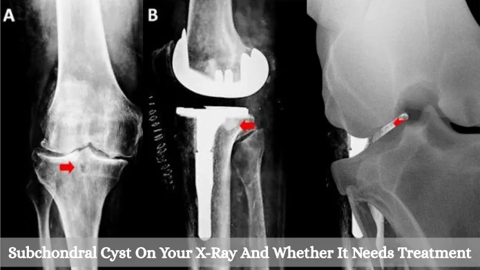
“Cyst” isn’t quite the right word here, and plenty of radiologists will tell you so themselves. What they spotted on your X-ray is a small hollow in the bone, sitting right under the cartilage of the joint, and it doesn’t have the lining that a true cyst would have. That is why some reports call it a geode instead, after the hollow rocks people crack open at gem shows.
Either name points to the same thing. For most people, a subchondral cyst will be a side effect of arthritis in that joint, and it won’t need any treatment aimed at it directly. What it can tell your doctor about the joint is a different matter, and that part gets less attention than it should.
What A Subchondral Cyst Looks Like On An X-Ray
On the film, you would see a dark round or oval patch inside the bone, close to the joint surface, with a thin white line around the edge. The dark patch is the hollow. The white line is bone that has thickened around it over time.
They’re usually small, often a few millimeters, though some grow to a couple of centimeters. You can have one or a small cluster in the same spot. The knee, the hip, the shoulder and the joint at the base of the thumb are where they turn up most.
These are not tumors, and they won’t spread to other parts of the body.
Why The Bone Hollows Out Under Worn Cartilage
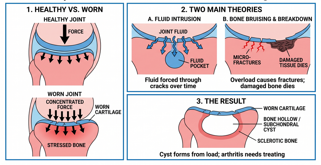
Healthy cartilage works like padding. It takes the force of each step and spreads it over the bone underneath. Once it wears thin, the bone right below starts taking more of that force than it was made to handle, and that is where the trouble begins.
There are two main theories on what happens next, and the experts seem to think both play a part:
- Fluid gets pushed in. Joint fluid can be forced through cracks in the thinning cartilage and into the bone, a little more with every step. Over months, it may carve out a pocket.
- The bone gets bruised and breaks down. Bone that is overloaded can pick up tiny fractures. The damaged patch may then die back and leave a gap behind.
So the cyst comes from the load on the bone. The arthritis is still the thing that needs treating.
Which Joint Problems Usually Come With It
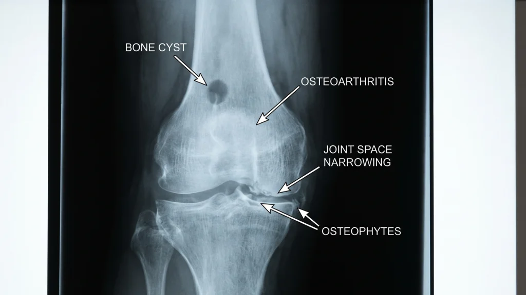
Osteoarthritis:
This will be the cause in the large majority of cases. If the same report mentions “joint space narrowing” (the cartilage has thinned) and “osteophytes” (bone spurs), the cyst belongs to that same picture of a worn joint.
Other Causes:
Less often, the cyst comes from something else, and your doctor will want to rule these out because they’re treated differently:
- Rheumatoid arthritis, where inflammation of the joint lining eats into the nearby bone.
- Pseudogout (CPPD), where calcium crystals build up in the cartilage.
- Avascular necrosis, where part of a bone loses its blood supply, most often at the top of the thigh bone in the hip.
- An old injury, such as a fracture that ran into the joint years ago.
Is The Cyst Causing Your Pain?
In most cases, probably not on its own. Lots of people have one and never feel a thing, and many are only found because a sore joint was X-rayed for some other reason. The morning stiffness, the ache on the stairs and that grinding feeling usually come from the arthritis as a whole.
It gets more complicated once an MRI is done. Knees with a cyst nearly always have a bone marrow lesion around it as well, which is an area of swollen, irritated bone. Those lesions are linked with pain. So while the cyst itself may be quiet, it often sits in the middle of bone that is sore.
When A Cyst Can Cause Trouble Directly
Every now and then, a cyst will grow large enough that the shell of bone above it gets very thin. If that shell cracks or sinks, you’ll likely notice the pain change. It tends to come on fast, it’s sharp when you put weight on the joint, and resting won’t ease it the way it eases a normal arthritis flare. If that happens, call your doctor’s office that week rather than waiting for your next visit.
Does A Cyst Mean The Arthritis Is Getting Worse?
It can, and this is the main reason your doctor will care about it.
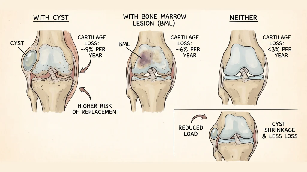
A group of Australian researchers followed people with painful knee arthritis for two years with MRI scans, and close to half of them had at least one subchondral cyst. This is what turned up when they compared the knees:
- Cartilage on the inner side of the knee wore away at about 9% a year in knees with a cyst.
- In knees with only a bone marrow lesion, it was closer to 6% a year.
- In knees with neither, it was under 3% a year.
- Over four years, a cyst on the inner side of the knee went along with higher odds of needing a knee replacement.
That sounds grim at first, but the same study had better news too. Cysts didn’t only grow. About 1 in 9 people saw a cyst shrink over the two years, and some disappeared completely. In those knees, less cartilage was lost.
Surgeons who straighten bowed legs have reported something similar. After the leg is realigned and weight moves off the worn side of the knee, the cysts on that side tend to get smaller.
The cyst seems to follow the load on the joint, so anything that takes pressure off the joint will also be working on the cyst.
How A Subchondral Cyst Is Treated
There isn’t a pill or a shot made for the cyst. Treatment goes after the arthritis and the stress on the joint, and the cyst is expected to settle along with it.
Exercise And Weight
If you only do one thing, strengthening the muscles around the joint would be the one to pick. For a knee or hip, that means the thigh and hip muscles. Stronger muscles take in more of each step, which leaves less for the bone. A physical therapist can set up a program that won’t set off a flare, and most insurance plans cover a course of visits with a referral.
Weight loss helps the knee in particular, since each pound you carry puts several pounds of force through the knee as you walk. Even a modest loss will take real pressure off.
Medicines And Injections
For day-to-day pain, the usual options are:
- Acetaminophen (Tylenol).
- An anti-inflammatory such as ibuprofen (Advil) or naproxen (Aleve), if your doctor says it’s safe for you.
- Diclofenac gel (Voltaren), rubbed onto a sore knee or hand, which is easier on the stomach.
- Ice or heat during a flare.
- A cane in the hand opposite the sore side, or an unloader brace for a knee worn mostly on one side.
A steroid injection into the joint can calm a flare for a few weeks or months. It works on the inflamed joint lining rather than the cyst, and most doctors will limit how many you get in a year. Draining a cyst with a needle is done now and then for a large one that keeps growing, but it isn’t routine.
Surgery
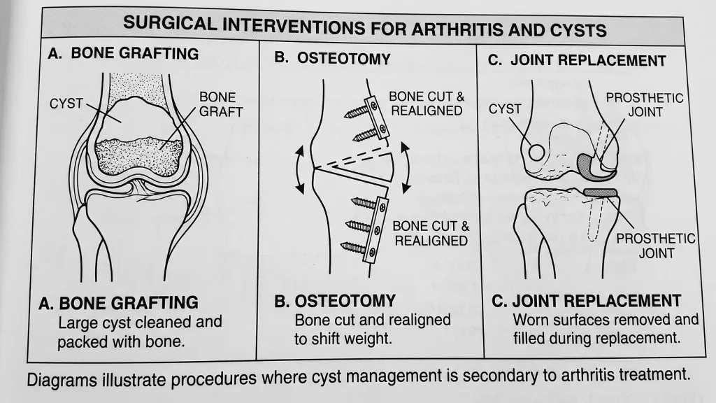
A small cyst is not a reason for surgery. When an operation does happen, it’s for the arthritis, and the cyst is dealt with along the way:
- Bone grafting, where a large cyst that threatens the joint surface is cleaned out and packed with bone.
- Osteotomy, where the bone is cut and realigned to move weight off the worn side, mostly in younger, active people.
- Joint replacement, where the worn surfaces are removed (cysts included) and any larger hollows are filled with graft during the same operation.
When The Spot On The X-Ray Needs Another Scan
Radiologists see these cysts every day and rarely confuse them with anything else. Once in a while, though, a hollow near a joint turns out to be a benign bone tumor or, much less often, cancer that has spread from somewhere else. If the radiologist isn’t sure, the report will normally say so and suggest an MRI or CT.
You should ask about a second scan if:
- The report calls it a “lytic lesion” or recommends more imaging.
- There is little or no arthritis in the joint, yet the spot is there anyway.
- The pain is deep and constant and gets worse at night or at rest.
- You’ve lost weight without trying or have had cancer before.
- The spot is large, has a ragged edge, or has grown since an earlier X-ray.
If none of those apply and the joint shows the usual signs of wear, the cyst is simply part of the arthritis. The conversation with your doctor should be about the joint as a whole and what can be done to take the load off it.